XROMM technology, or X-ray Reconstruction of Moving Morphology, gives researchers a look at bones in motion. This could not only have an impact on human medicine, but also further science’s understanding of animal anatomy. And as if this isn’t enough motivation to advance this technique, the scientists at Brown University also believe that the field of biomechanics could be transformed by it.
###moving-bones###
The scientists make use of XMALab, a new open-source software package. With using open-source software, the scientists make sure that XROMM is accessible to other users.
Combining X-rays and CT scans
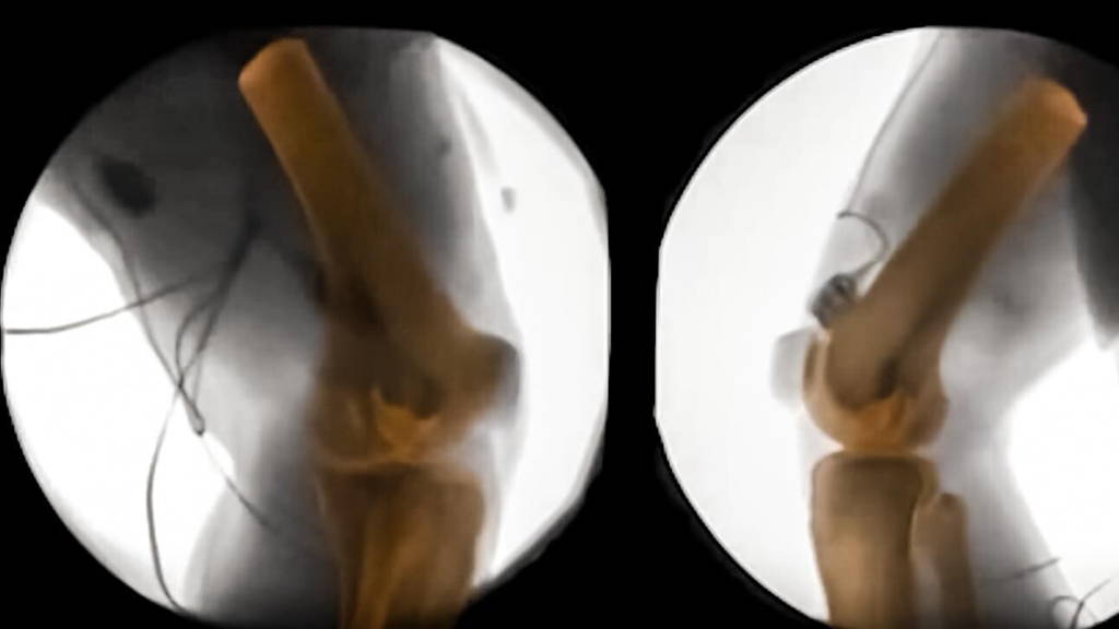
X-ray machines have been giving insight into the human body for quite some time now. X-rays can even show movement in unprecedented detail, like blood flowing through a body. One thing such machines can’t provide however, is a 3D-image. Enter CT scans. These scans show detailed, high-quality images of the body in 360 degrees. The scientists at Brown decided to layer these two scans, which led to the XROMM technology and a look at bones in motion.###moving-bones###
Understanding how bones move
The new technology helps scientists understand bones more. It, for example, shows how the shape of bones relates to the way the bones move. It also gives scientists a better look at how feet touch the ground, or why prostheses are deemed uncomfortable by amputees. Collaborators at the VA hospital in Providence are already looking at how the residual limb of an amputee moves inside a prosthesis, to try to improve the interface. It could also provide insight as to why women suffer more knee injuries than men. The possibilities seem endless, for humans and animals alike.The scientists make use of XMALab, a new open-source software package. With using open-source software, the scientists make sure that XROMM is accessible to other users.