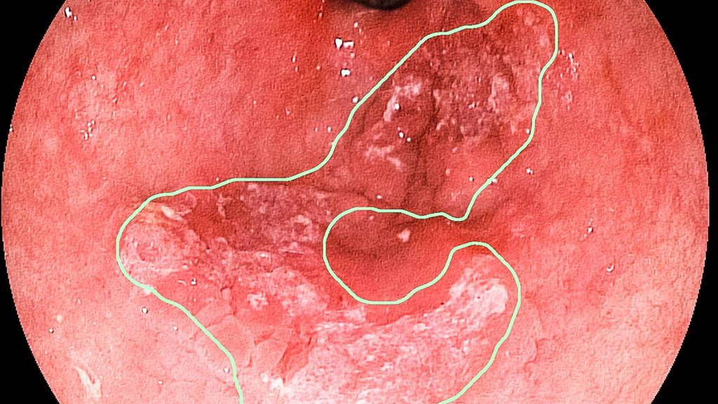
People with long-lasting reflux often develop irritation because of the gastric acid, which can lead to abnormal tissue in the esophagus. These so-called Barrett's esophagus is one of the main risk factors for developing esophageal cancer in the Western world. An endoscopic control is one of the methods to see if people have the disease. But the earliest forms of cancer are very difficult to spot and missed regularly. The survival rate for people with advanced cancer is only 50 percent. The number of people with reflux increases, because it is more common in overweight people. And there are more and more overweight people.
Gastroenterology and Liver physician Dr. Erik Schoon of the Catharina Hospital in the Netherlands has teamed up with the Video Coding and Architectures Research Group at the TU / e. This group, led by Professor Peter de With, has considerable experience with image analysis techniques to recognize objects and people in for example smart cameras.
TU/e researchers Fons van der Sommen and Sveta Zinger, both from the VCA, developed together with dr. Erik Schoon new techniques to studie the endoscopic images on the early signs of cancer. These techniques are now so good that the recognition score rivals the best European specialists. "Spectacular", Schoon says about the results. "Recognizing early cancer in Barrett's esophagus is one of the hardest things in our profession."
The research is conducted by the Eindhoven University of Technology in collaboration with the Catharina Hospital in Eindhoven, University Hospital Leuven, the Krankenhaus Barmherzige Brüder Regensburg, St. Antonius Hospital Nieuwegein and the Academic Medical Hospital in Amsterdam. The publication in the leading scientific journal Endoscopy, entitled Computer-aided detection of early neoplastic lesions in Barrett's esophagus "can be found at DOI number 10.1055 / s-0042-105284.
Gastroenterology and Liver physician Dr. Erik Schoon of the Catharina Hospital in the Netherlands has teamed up with the Video Coding and Architectures Research Group at the TU / e. This group, led by Professor Peter de With, has considerable experience with image analysis techniques to recognize objects and people in for example smart cameras.
TU/e researchers Fons van der Sommen and Sveta Zinger, both from the VCA, developed together with dr. Erik Schoon new techniques to studie the endoscopic images on the early signs of cancer. These techniques are now so good that the recognition score rivals the best European specialists. "Spectacular", Schoon says about the results. "Recognizing early cancer in Barrett's esophagus is one of the hardest things in our profession."
Much less intrusive
Computer analysis should eventually be available in all hospitals, to help gastroenterologists in recognizing the earliest stages of cancer. That figure would be recognized in time and treated cancer can toward 100 percent. Many patients wouldn't need surgery which removes a portion of the esophagus. Often unavoidable at late discovery. Treatment of early cancer is much less invasive for patients, and usually consists of a micro-surgical procedure from the inside. It is also much cheaper. In addition, physicians who are not specialized in Barrett, defects learn to recognize more quickly with the aid of this technique.Realtime
Before the new technique is applied, the software still needs to be improved and adapted to analyze real-time video image, and then will follow extensive testing in hospitals. It will probably take another five to ten years before it can be widely introduced. Last week it was announced that will be made available half a million euros for further research, including KWF and STW.The research is conducted by the Eindhoven University of Technology in collaboration with the Catharina Hospital in Eindhoven, University Hospital Leuven, the Krankenhaus Barmherzige Brüder Regensburg, St. Antonius Hospital Nieuwegein and the Academic Medical Hospital in Amsterdam. The publication in the leading scientific journal Endoscopy, entitled Computer-aided detection of early neoplastic lesions in Barrett's esophagus "can be found at DOI number 10.1055 / s-0042-105284.
