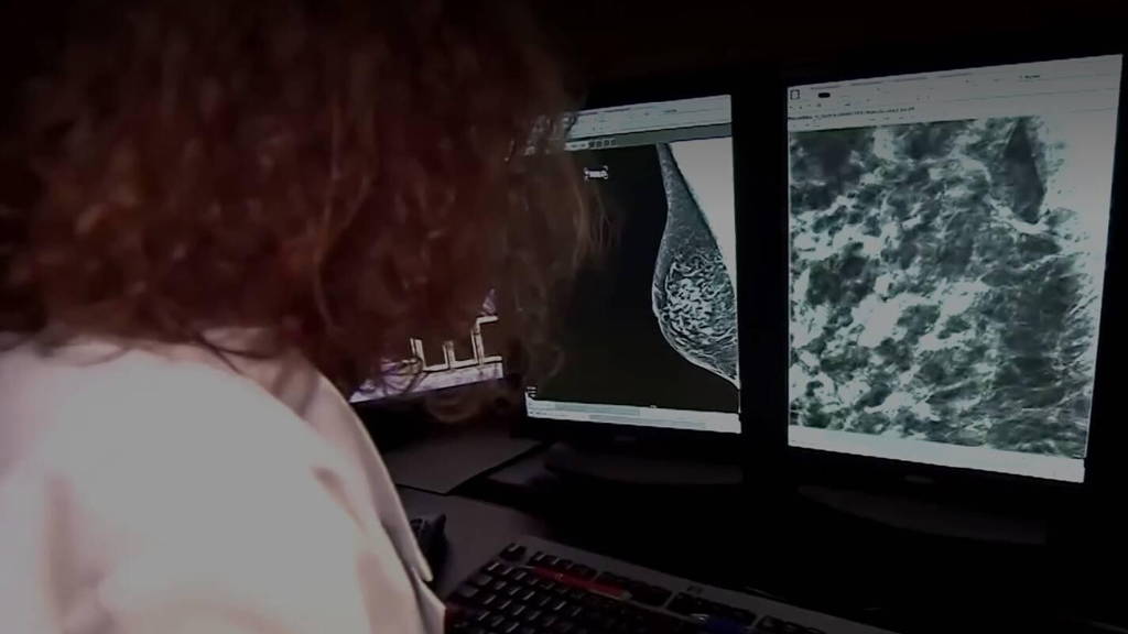
The use of AI can significantly improve the quality of breast cancer screening, research shows. Improving risk predictions of breast cancer can also improve its prevention. Improved techniques with AI make it easier to see on mammograms what the mammographic density, also known as breast tissue density, is. This is important in screening for breast cancer.
Breast tissue that looks white on a mammogram is radiologically dense, while breast tissue that looks dark is considered non-dense. In general, it can be said that women who have a higher mammographic density for their age and body-mass index also have a higher risk of breast cancer. On top of that, in the case of higher density, it is more difficult to detect breast cancer through mammography. The study was recently published in the journal Trends in Cancer.
Leading the way for further research
Mammographic density in some cases guides the use of additional imaging technologies. Examples include ultrasound and magnetic resonance imaging (MRI) that provide increased cancer detection rates in clinical trials in women with extremely dense breasts. To predict a future diagnosis of breast cancer, technology such as deep learning is already being used to analyze mammographic images.
A major advantage of this is that AI can uncover mammographic features that are strong predictors of breast cancer risk. This can also better explain the relationship between mammographic density and breast cancer risk. But scientists and researchers continue to struggle to find a good explanation in the relationship between the two, mammographic density and breast cancer risk. “A woman with mammographic features associated with a high risk of breast cancer detection may benefit from more frequent screening or risk-reducing medication,” said lead researcher and author, Erik Thompson, of Queensland University of Technology in Brisbane, Australia.
Additional imaging
In doing so, he also says that a woman with a low chance of being diagnosed with breast cancer could have a longer interval between screenings over the next five years. In addition, a woman with high mammographic density without high-risk mammographic features could benefit from additional imaging, such as MRI or ultrasound.
Still, according to the study, it does not always remain clear when AI identifies mammograms as cancer or not. “It is critical that we identify the pathobiology associated with mammographic features and the underlying mechanisms that link them to the genetic, inherited predisposition to breast cancer,” Thompson says.
Improve breast cancer screening
The fact is that high breast tissue density is associated with an increased risk of developing breast cancer. Adding to that, as already reported, breast cancer screening is complicated by breast tissue density. There are several approaches being explored on how to improve such screening. For example, researchers at the University of Eastern Finland developed a new artificial intelligence-based algorithm, MV-DEFEAT, to improve the assessment of mammographic density.
Researchers at the University of Edinburgh recently developed another AI algorithm that makes it possible to detect breast cancer at an even earlier stage. With the developed AI algorithm combined with the non-invasive laser analysis technique, Raman spectroscopy, subtle changes in the bloodstream can be recognized.


