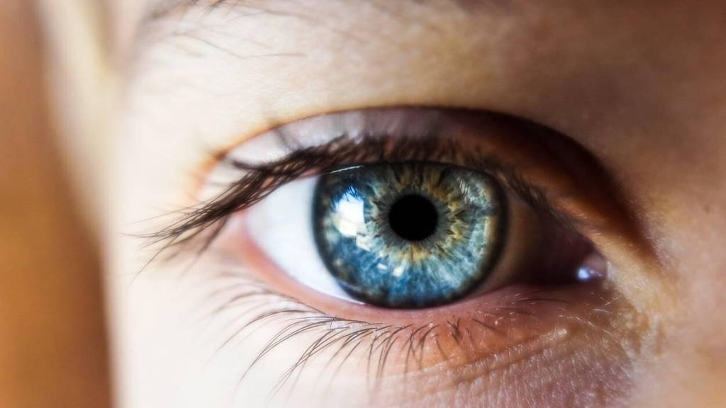
People with diabetes have a higher risk of damage to the blood vessels in the retina. This can lead not only to impaired vision, but in some cases even blindness. This is why diabetic patients have to visit the ophthalmologist regularly for check-ups. There, a fundus photograph is then taken to map and examine the retina's blood vessels and determine whether the patient suffers from diabetic retinopathy. A process that sometimes takes three quarters of an hour. A newly developed AI tool can speed up this examination and improve diagnostic accuracy.
To examine a fundus photo, ophthalmologists currently have to manually segment the retinal image. This involves distinguishing between the background and blood vessels of different length, width and swelling pattern. Apart from diabetic retinopathy, this method is also used to look for other eye and cardiovascular diseases. This manual segmentation process is not only very difficult, but also time-consuming and error-prone.
AI tool speeds up and improves diagnostics
Researchers at a joint laboratory of Sharjah's Skoltech University and the AIRI Institute have developed an AI tool for this manual and time-consuming task. This allows automated analysis of retinal photos used to diagnose diabetic retinopathy. This speeds up the whole diagnosis process, and also allows diagnoses to be made more accurately.
There are already several other examples of AI technology being used to analyse eye photos and diagnose (eye) conditions. For instance, back in 2021, a method was developed to analyse fundus photos for the presence of glaucoma using an AI algorithm. Last year, researchers at the Johns Hopkins Children's Centre discovered that an AI-driven eye examination of young people with diabetes could increase the chances of early detection of diabetic eye diseases.
High accuracy as well as sensitivity
An initial test on three sophisticated datasets achieved an accuracy of 97 per cent. According to the researchers, however, that is not the most important result. The sensitivity of the AI tool analyses came out at 84 per cent, based on the industry-standard DRIVE database. ‘Sensitivity is the most important thing. It reflects the model's ability to identify micro vessels. Something that previous models struggled with,’ said Melaku Getahun, a Skoltech PhD student and lead author of the study.
What makes this kind of segmentation particularly challenging are the fine details in retinal images, which must be taken into account and yet often escape both the neural networks previously developed for this task and some eye specialists who analyse these images manually
Challenges
Although the initial results can be called promising, the researchers still have some challenges to overcome. For instance, the datasets used to train and test the AI model still had some limitations, especially in terms of size. ‘This hampered the model's ability to effectively generalise to unseen data. However, by carefully applying data augmentation and processing techniques, we managed to significantly improve the model's performance,’ said the study's principal investigator. ‘Even with our new neural network architecture, the problem remained that certain pixels of microvessels were misclassified as background. To address this, we implemented an adaptive threshold algorithm, which yielded a significant improvement in sensitivity and accuracy.’
Future prospects
These challenges do not mean that the AI tool developed for analysing retinal photographs does not have a future. Its ability to recognise tiny unhealthy blood vessels could certainly be of value for clinical use. If the aforementioned challenges are overcome and the system is properly further developed, the researchers say it could become a standard tool for screening for eye diseases, allowing ophthalmologists to diagnose conditions faster and more accurately.
‘This could help in the early diagnosis and prevention of hard-to-treat eye diseases, such as diabetic retinopathy, which is common in populations with high diabetes incidence, as well as other related microvascular eye diseases,’ added co-author of the study and professor Rifat Hamoudi from the University of Sharjah. The research was recently published in Pattern Recognition Letters.


