To understand how it works, Quartz Media offers the example of tuberculosis treatment. When a patient in, say, rural India is diagnosed with the disease, a doctor without access to a sophisticated lab will typically give the patient drugs from the first line of treatment. However, one in four patients won’t respond to this treatment, because the bug they’ve caught is resistant to these drugs.
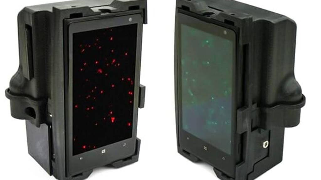
Without a test to determine which strain the patient is suffering from, the doctor has to wait for weeks till he or she finds out that the patient has the drug-resistant infection, and then prescribe a different medication. In that period, the patient would have already spread the infectious and hard-to-treat disease to many more people around him. A portable DNA analyzer could easily fix this problem. To create one such analyzer, Mats Nilsson of Stockholm University and his colleagues took a sample of human tissue and analyzed it with a specially designed attachment connected to a Nokia Lumia 1020 smartphone with a special 41 megapixel camera.
Next, a new set of chemicals, which become fluorescent when light is shone on them, attach themselves to it based on what the sequence is. Finally, the attachment shines two different colors of laser onto the mixture. With all those DNA snippets emitting light, a decent smartphone camera is now able to see DNA. If the tuberculosis strain is drug-resistant, the mixture shines in a different color than if the infection is not drug-resistant. All this can happen in the space of a few hours, rather than the days or weeks that would be needed to send the sample to a specialized lab for the same result. “I was so surprised,” says Nilsson, “the images from the smartphone and those from the lab were indistinguishable.”
There are limitations, though. The lab has yet to develop a method that engineers the movement of various liquids that go in and out of the attachment. To show their system works, the researchers used lab equipment to do it. And at present, the system can only test for one type of disease at a time. So if you want to test for cancer after a tuberculosis test, you’d have to wash the equipment and use a whole different set of chemicals to perform the analysis.
Nilsson thinks it should be possible to adapt the attachment for different smartphones, even those with lower-resolution cameras. The Nokia Lumia 1020 has a special 41-megapixel camera, where typical smartphone cameras in the latest generation use 12 megapixels.
The project also faces competition. A UK-based company, Oxford Nanopore, has developed a technology that doesn’t rely on cameras to achieve a similar analysis. It is in the process of creating a smartphone adapter for it. The system promises to be even more portable, because it won’t require the use of specialized chemicals. Clive Brown, the company’s chief technology officer, told the BBC that it would “allow anybody to sequence anything, anywhere.”
The study’s results were published in Nature Communication.
Without a test to determine which strain the patient is suffering from, the doctor has to wait for weeks till he or she finds out that the patient has the drug-resistant infection, and then prescribe a different medication. In that period, the patient would have already spread the infectious and hard-to-treat disease to many more people around him. A portable DNA analyzer could easily fix this problem. To create one such analyzer, Mats Nilsson of Stockholm University and his colleagues took a sample of human tissue and analyzed it with a specially designed attachment connected to a Nokia Lumia 1020 smartphone with a special 41 megapixel camera.
Tiny section genome analysed
DNA is made up of four bases: A, T, G, and C. A tuberculosis genome consists of typically 4.5 million base pairs. But Nilsson only needs to look at a tiny section of the whole genome. Inside the attachment, specialized chemicals are designed to seek that tiny sequence of DNA and snip it out. Since DNA is too small for any smartphone cameras to capture, an enzyme is used to multiply the small snippet 1,000 times.Next, a new set of chemicals, which become fluorescent when light is shone on them, attach themselves to it based on what the sequence is. Finally, the attachment shines two different colors of laser onto the mixture. With all those DNA snippets emitting light, a decent smartphone camera is now able to see DNA. If the tuberculosis strain is drug-resistant, the mixture shines in a different color than if the infection is not drug-resistant. All this can happen in the space of a few hours, rather than the days or weeks that would be needed to send the sample to a specialized lab for the same result. “I was so surprised,” says Nilsson, “the images from the smartphone and those from the lab were indistinguishable.”
Fraction of the cost
The study’s results were published in Nature Communications. Nilsson’s team is now looking to commercialize the technology. The researchers believe that the attachment could cost as little as $500, a fraction of the many thousands of dollars needed for lab equipment.There are limitations, though. The lab has yet to develop a method that engineers the movement of various liquids that go in and out of the attachment. To show their system works, the researchers used lab equipment to do it. And at present, the system can only test for one type of disease at a time. So if you want to test for cancer after a tuberculosis test, you’d have to wash the equipment and use a whole different set of chemicals to perform the analysis.
Nilsson thinks it should be possible to adapt the attachment for different smartphones, even those with lower-resolution cameras. The Nokia Lumia 1020 has a special 41-megapixel camera, where typical smartphone cameras in the latest generation use 12 megapixels.
The project also faces competition. A UK-based company, Oxford Nanopore, has developed a technology that doesn’t rely on cameras to achieve a similar analysis. It is in the process of creating a smartphone adapter for it. The system promises to be even more portable, because it won’t require the use of specialized chemicals. Clive Brown, the company’s chief technology officer, told the BBC that it would “allow anybody to sequence anything, anywhere.”
The study’s results were published in Nature Communication.